OCT Scanning
Optical Coherence Tomography. Non-invasive, painless, and capable of detecting eye disease before symptoms appear.
What is OCT?
Optical Coherence Tomography is a non-invasive imaging test. OCT uses light waves to take cross-section pictures of your retina. With OCT, your optometrist can see each of the retina's distinctive layers and map and measure their thickness.
These measurements help with the diagnosis of conditions such as glaucoma and macular degeneration, often detecting changes before any symptoms are noticed.
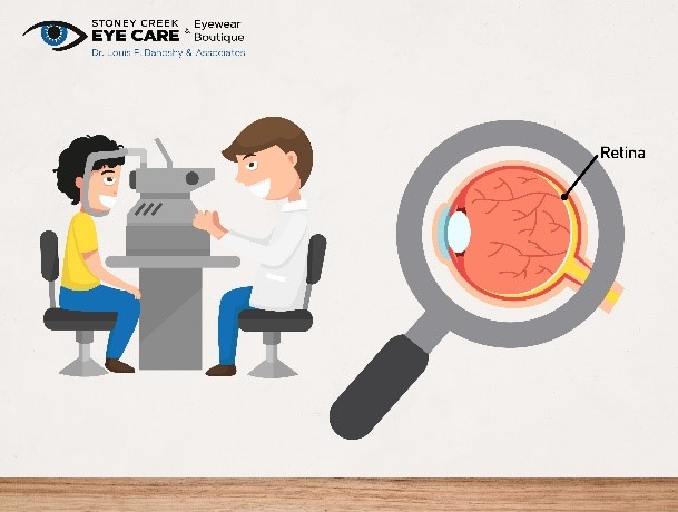
How It Works
The test takes about 10 minutes to complete. It is non-invasive and entirely painless. You place your chin on a rest and keep your eyes open as you look at a small light within the machine. The OCT machine scans your eye while you hold still.
Fundus Photography vs OCT
We carry out fundus photography with each examination, currently at no charge. This provides a standard photograph of the back of your eye.
OCT scanning goes further. It produces a detailed cross-section of the retinal layers, allowing us to detect subtle changes that a standard fundus photograph would not reveal. OCT is not done as standard but is available upon request.
Conditions OCT can help detect:
- Glaucoma
- Age-related macular degeneration (AMD)
- Diabetic retinopathy
- Macular holes and epiretinal membrane
- Vitreous detachment
Request OCT Scanning
Ask about OCT scanning when booking your next eye examination, or call us to find out more.